Endoscopic spine surgery · Louisville, KY

UBE spine surgery

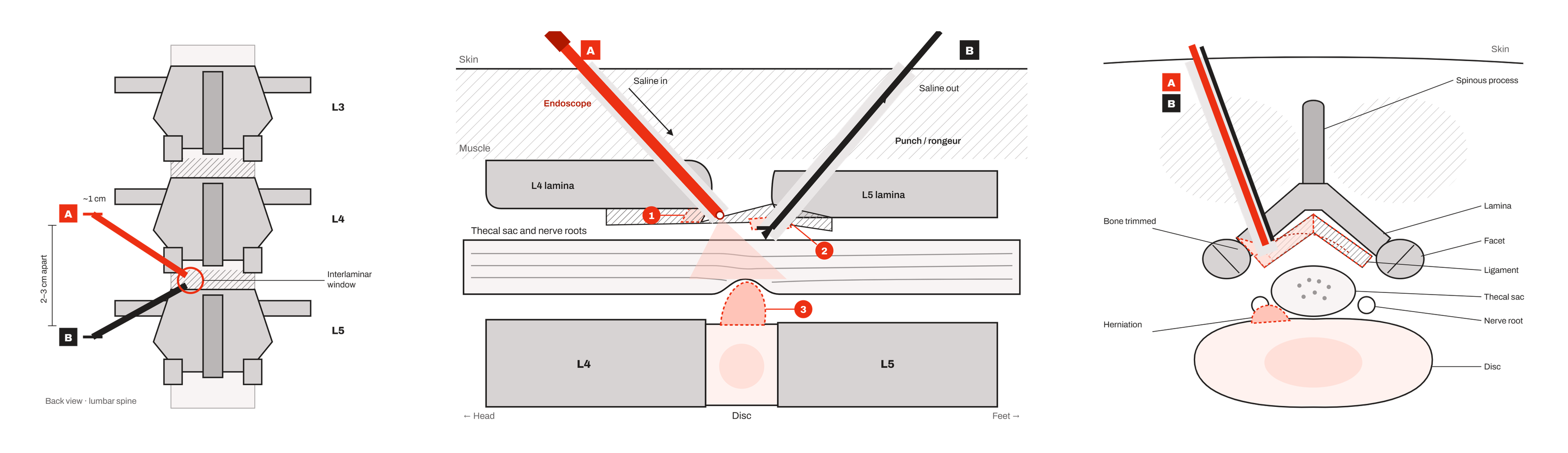
Three diagrams of UBE spine surgery: back view of the two portals at the interlaminar window, side view of the endoscope and instrument inside the canal reaching a disc herniation, and a cross-section showing the small amount of bone trimmed beside the nerve root

Ask whether UBE is an option for your back.

Request an appointment

Two small openings. One camera. The least tissue we can disturb.

UBE stands for unilateral biportal endoscopic surgery. In plain terms: instead of one incision, Dr. Vemuri uses two openings each smaller than a fingertip. A camera goes through one and the instruments through the other, with a gentle stream of saline keeping the view clear. It lets him free a pinched nerve or remove a disc fragment while disturbing less muscle than a standard approach. Most UBE cases go home the same day. No referral needed, and same-week appointments are available. Hablamos español: (502) 362-1780.

Call (502) 242-6370
2
Small openings, not one incision
0
Nights in the hospital for most UBE cases
No
Referral needed
5,000+
Spine surgeries by Dr. V
2
Small openings, not one incision
0
Nights in the hospital for most UBE cases
No
Referral needed
5,000+
Spine surgeries by Dr. V

How the surgery goes.

Two small openings

Dr. Vemuri makes two openings, each smaller than a fingertip, on one side of the spine. No muscle is cut away from the bone.

Camera in one, instruments in the other

The endoscope gives a magnified view on a screen. Under saline, the bone spur, ligament or disc fragment pressing on the nerve is removed.

Closed with a stitch or two

The openings are small enough to close with a stitch each. Most UBE patients at miiSpine go home the same day.

What UBE is.

Standard minimally invasive spine surgery works through one tube with a microscope looking down it. UBE uses two separate openings a short distance apart. An endoscope, a thin camera, goes through one, and the working instruments go through the other. Saline flows through the whole time, which keeps the view clear and clean. Because the camera sits right next to the nerve, Dr. Vemuri sees the anatomy up close and from an angle a microscope can't reach, through openings measured in millimeters. UBE has been used internationally for more than a decade for disc herniations and stenosis in the lower back.

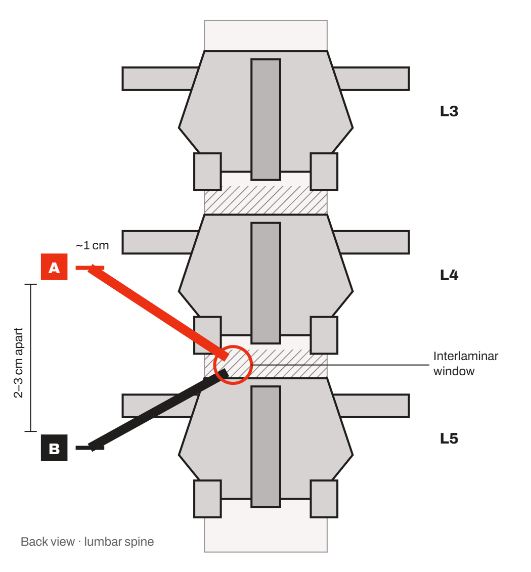
Diagram, back view of the lumbar spine: two UBE portals, each about 1 cm and 2 to 3 cm apart, meeting at the interlaminar window between L4 and L5

Back view. Two openings about 1 cm each, 2 to 3 cm apart, meet at the small window between two laminae. A is the camera, B is the working instrument.

What to expect.

Before

A visit with Dr. Vemuri and your MRI reviewed with you in the room. He'll tell you whether UBE, a standard minimally invasive approach, or no surgery is the right call.

The day of surgery

UBE at miiSpine is usually outpatient surgery. You arrive in the morning, have the procedure, spend a few hours in recovery, and go home.

After

Walking starts the same day. You get a written plan for activity, driving and work. Everyone heals at their own pace, so the plan is yours, not a generic timeline.

When UBE makes sense, and when it doesn't.

UBE is a way of doing a decompression or a microdiscectomy, not a reason to have one. The decision to operate is the same as always: conservative care first, surgery when it hasn't worked or when there's real weakness. Once surgery is the plan, UBE suits many lower-back disc herniations and stenosis cases, including some repeat herniations after a previous operation. It isn't the right tool for every spine. Dr. Vemuri does standard minimally invasive surgery too, so he picks the technique that fits your anatomy rather than the one with the newest name.

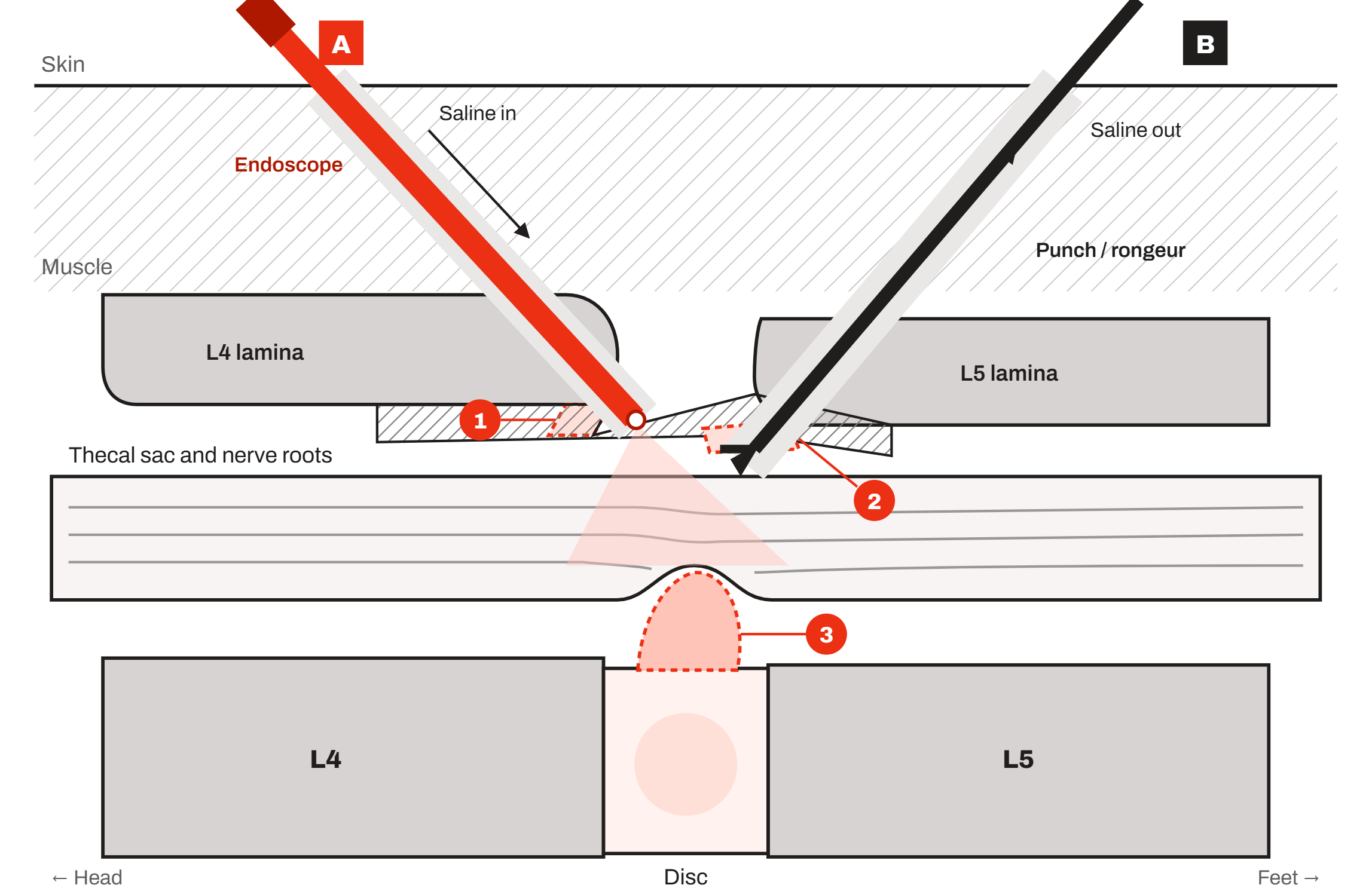
Diagram, side view inside the spinal canal: the endoscope and a working instrument enter between the L4 and L5 laminae, with saline flowing in and out, to reach a disc herniation pressing on the nerve roots

Side view. The camera (A) and the instrument (B) pass between the L4 and L5 laminae. Saline flows in through one portal and out the other, keeping the view clear. 1 and 2 are the small amounts of bone and ligament trimmed to open the window; 3 is the disc herniation being removed.

What UBE can treat.

A herniated disc in the lower back

A disc fragment pressing on a nerve and causing leg pain, numbness or weakness, including some herniations that have come back after a previous surgery.

Spinal stenosis

A narrowed canal that causes leg pain or cramping with walking. UBE can open the canal while leaving the bone and ligament that aren't in the way.

A pinched nerve where it exits the spine

Narrowing of the small opening a nerve passes through, called foraminal stenosis, which causes persistent buttock or leg pain.

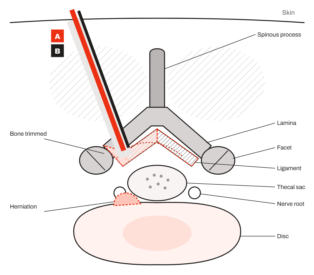
Diagram, cross-section of a lumbar vertebra: the two UBE instruments reach the disc herniation beside the thecal sac and nerve root after a small amount of bone and ligament is trimmed; the facet joints are left alone

Cross-section. Both instruments come in from one side. A small edge of bone and ligament is trimmed to reach the herniation next to the nerve root. The facet joints and the muscle on the far side are left alone.

5,000+
Spine surgeries
4,000+
Office visits per year
1,000+
Outpatient procedures in five years

Dr Venu
Vemuri

Dr. Venu Vemuri, D.O. (Dr. V) is a fellowship-trained spine surgeon and is board-certified by the American Osteopathic Board of Orthopedic Surgery. He opened miiSpine in 2021 as an independent alternative to corporate medicine. He has performed more than 5,000 spine surgeries, and he trained in UBE because, for the right patient, it disturbs less tissue than anything else he does.

View More ↗

Questions about UBE.

What does UBE stand for?

Unilateral biportal endoscopic surgery. Unilateral means both openings are on one side of the spine. Biportal means there are two of them. Endoscopic means a thin camera does the looking.

How is it different from standard minimally invasive surgery?

Standard minimally invasive surgery works through one tube with a microscope. UBE uses two smaller openings, a camera that sits next to the nerve, and saline to keep the view clear. In our experience that means smaller openings and less muscle disturbance for the cases it suits.

Is UBE outpatient surgery?

Usually, yes. Most UBE cases at miiSpine go home the same day.

How long does recovery take?

It depends on what was treated and on you, so we don't promise a date. Walking starts the day of surgery, and Dr. Vemuri gives you a written plan for activity, driving and returning to work.

Am I a candidate?

The only way to know is for Dr. Vemuri to look at your MRI with you. UBE suits many lower-back disc herniations and stenosis cases. It isn't the answer for instability or for every neck problem.

¿Hablan español?

Sí. Llame al (502) 362-1780, las 24 horas, o visite miispine.com/accidentes.

Is UBE new?

It has been used internationally for more than a decade, with a growing body of published research. It's newer to the United States, which is why fewer surgeons here offer it.

Do I need a referral?

No. Call (502) 242-6370 or request an appointment online. Same-week appointments are available.

Does insurance cover UBE?

Coverage depends on your plan and on what's being treated. Our team checks your benefits before surgery and tells you what to expect. If you're paying cash, we'll give you the price before you book.

This content is for general information and isn't a substitute for a medical evaluation. Every spine is different. Talk to a spine specialist about your specific symptoms.

Our latest studio
news & insights.

A team of programming experts and designing
professionals develops, design, runs test, and/or manages
software development for the below-written :